150 shades of gray
Text: Arthur Toporkov, Lilia Reimbaeva, Pavel Severin

MRI «portraits» are precise depictions of our inner constitution |
According to estimates, the average human is able to see thirty different shades of gray. Some talented writers manage to detect up to fifty. But a decent medical radiologist makes the true champion: these professionals can tell apart about 150 shades! After all, it's part of their job. They practice daily at work, thanks to the images MRI machines create that have a wide spectrum of gray shades. The ability to distinguish each shade is necessary for reaching correct medical conclusions, which can be literally vital when other examination methods fail.
Today the acronym MRI is familiar to more or less everyone. Sounding vaguely like the name of a secret service agency or a space technology and looking like a piece of arcane heavy machinery — which is basically what it is — this huge doughnut barely inspires confidence in a lay person, no matter how often Dr. House resorts to using it. However, MRI has proven its worth as a diagnostic technology that has already become indispensable in today's radiology centers. And here is why.
|
Ironically, the physics behind the MRI process were first explained in 1973 by a… chemist, American professor Paul Lauterbur. The Briton Peter Mansfield found a way to convert the obtained data into an image meaningful to the human eye (guess physicists know their stuff after all). Later they were awarded the Nobel Prize for their discoveries. Curiously, 13 years earlier,
in 1960, the Soviet scientist Vladislav Ivanov tried to patent the same
technology and similar equipment. To be precise, at the time he was not even a
|
How it works?
Magnetic resonance imaging, which is what MRI stands for and
what the doughnut essentially does, is a relatively recent invention. Its
operation is based on measuring the electromagnetic response of atomic nuclei —
typically of hydrogen — when exposed to a certain combination of
electromagnetic waves in a constant magnetic field… Okay, the scientific
We humans are a bit less than 80% water — that is, almost completely. Water, in turn, is a compound of hydrogen and oxygen with the ratio of two to one. So we have a whole lot of hydrogen in our body. Among other properties of this element, its particles can react to perturbations of a powerful magnetic field. And these reactions can be different depending on which body tissue the hydrogen particles belong to.
Essentially an MRI machine is just a large magnet. Usually, an electromagnet, although there are exceptions. This is why it is shaped like a doughnut, in fact: it contains an induction coil. This electromagnet has other devices attached to it, so not only the entire machine generates a magnetic field but also creates perturbations in it and can all the while record the reactions of the hydrogen particles contained in the object inside the field.
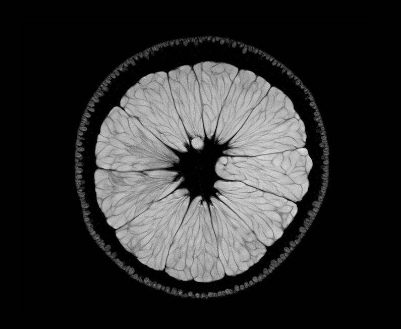
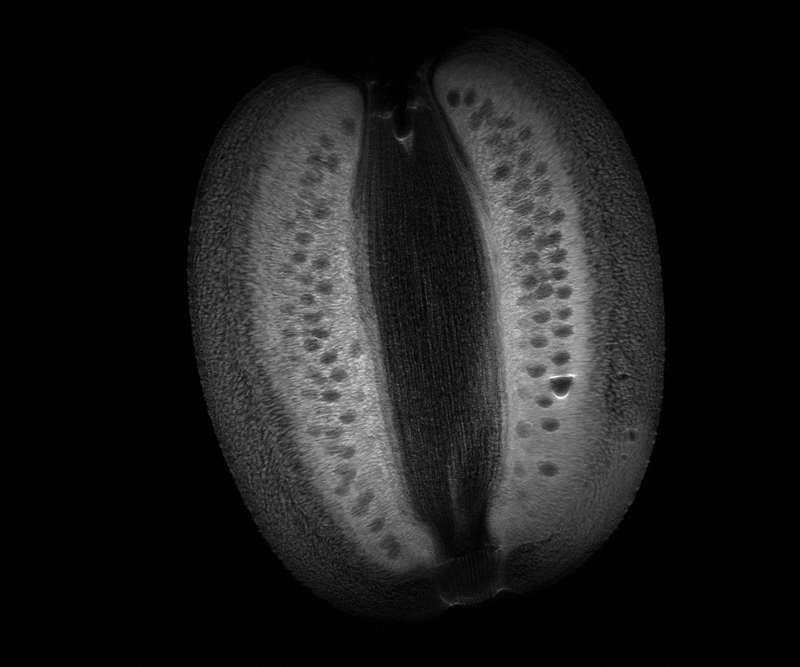
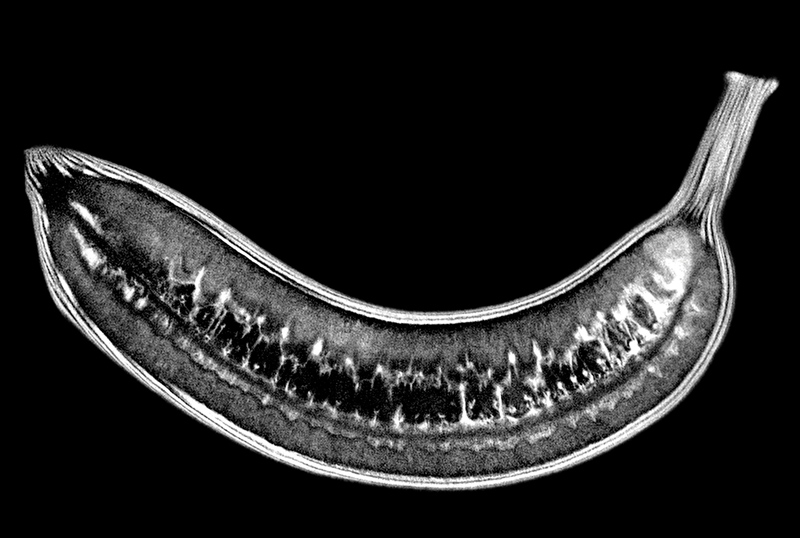
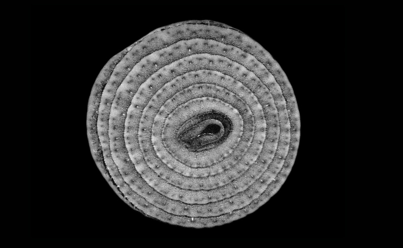
Usually the object is a human body, but if we place an orange inside the MRI scanner, we will get a detailed 3D−model of the fruit, which can be viewed from any angle and in any geometrical plane. Of course, for practical purposes we can easily find out what's inside an orange by taking a knife and slicing it open. But a human body can't be dissected so casually and certainly not in every spot and direction doctors need to look.
Thankfully, MRI makes it possible, «slicing» the patient electromagnetically in all planes at once and creating highly detailed scans of each «slice». The resulting images — which are many, so the process takes some time — are then combined into a single picture and… voila! you have a unique opportunity to literally look inside yourself. More importantly for diagnostic purposes, your doctor can take that look without having to invite you to the operating table.
Basically, an MRI scan is your interior selfie. And the doughnut, to continue the metaphor, is a highly sophisticated smartphone - a really large stationary one, and with a pretty good camera.
|
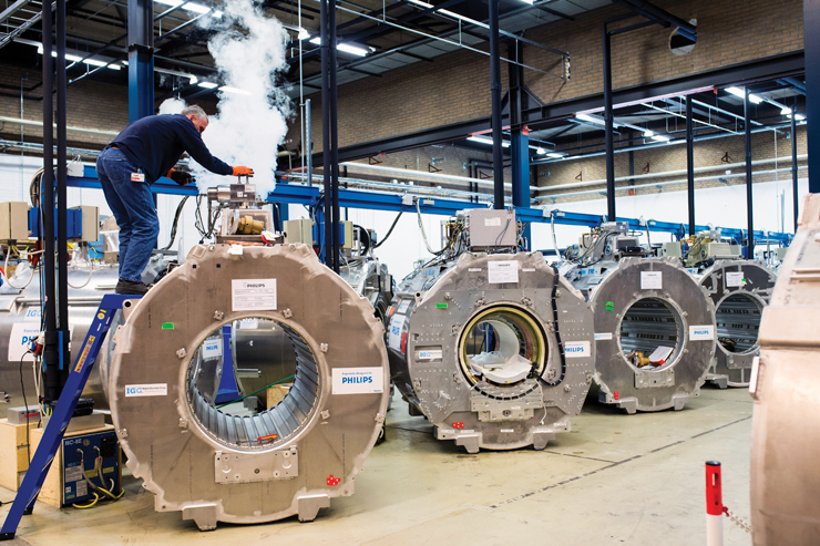
Behind the scenes: Philips medical equipment manufacturing division |
Why MRI?
One might say, «Those smartphones are a pricey pleasure… I
could take pictures
First off,
The point is, unlike MRI, which can be used any number of
times,
|
One of the most powerful MRI machines, 7 Tesla weighing 32 tons, is being transported |

Second,
Finally, in many cases MRI turns out to be the only way to produce accurate images of internal organs. There is a broad array of ambiguous signs pointing to a number of possible diagnoses. Headache, dizziness, motor dysfunction, and other symptoms may be a result of a minor fatigue or of a fairly serious condition. MRI is a primary instrument of modern differential diagnostics.
 |
What to look for?
Nowadays MRI is no longer a rare sight. While not necessarily every medical facility is equipped with an MRI machine, there is plenty of places where you can have a scan. If you need one, here is what you should look for when choosing a clinic.
Be sure to find out the magnetic flux density of the machine. It is called more commonly 'magnet strength' and measured in teslas (T). Ultimately, this number determines the MRI image quality. Going back to the smartphone metaphor, the tesla value can be compared with the number of megapixels. Most radiology centers are equipped with machines that have magnet strength of 1.5 T, while 3 T are somewhat less common — but either are enough for reliable diagnostics. MRI scanners with more than 3 T are used for scientific research purposes. But if you are told the scanner you intend to get examined with has less than 1.5 T (1 T, for example), it is better to look elsewhere: as the magnet strength decreases, the image quality falls exponentially.
Also, in any case bear in mind that an MRI machine cannot identify a medical condition. However advanced, this equipment is only a tool used by radiologists. It is their job to analyze the images, and their professional expertise enables them to interpret the scan results correctly.
Of course,
|
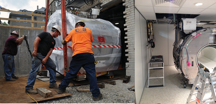
The photo on the left: Installing this kind of equipment requires complex engineering solutions The photo on the right: The new MRI machine is being installed in the Botkin City Hospital |
New MRI facility in Moscow
The choice of great radiology centers in Moscow is about to become wider. The Botkin City Hospital is currently being equipped with a new 1.5 T Philips MRI scanner complete with powerful modern software for a broad spectrum of diagnostic tests.
The entire project including planning and support is being accomplished by the MedImport company. In fact, the installation services being provided are a comprehensive, turnkey job, from assistance with selecting the scanner model to facility construction and equipment installation works.
To tell the truth, some of the tasks faced by MedImport have been quite peculiar. For example, in order to place the MRI doughnut inside, the construction team had to partially dismantle the building's outer wall — this was the only way to deliver the whopping 4.5−ton machine into the room where it would reside. As for the total weight of all equipment, it amounted to nearly 22 tons.
But where is all this, you would likely wonder upon visiting
the clinic. The fact is that the MRI machine — usually the only thing that the
patient sees in the scanning area — is only the tip of the iceberg. In order
for the MRI device to operate normally, an entire additional network of utility
lines had to be set up in the building. The cooling system alone has been a
project unto itself!
Instead of a conclusion, here is another trivia figure: the amount of helium used in the MRI cooling circuit would be enough to fill about four hundred thousand party balloons. And that many balloons is enough to lift a compact luxury car or… everyone's mood. Stay well!
|
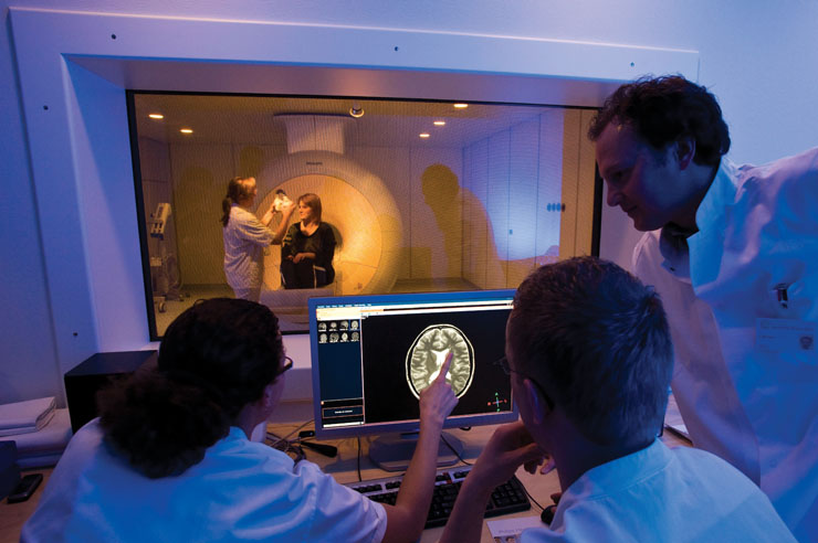
Often there is simply no alternative to an MRI examination |
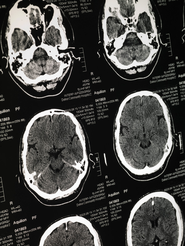
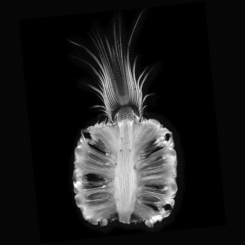
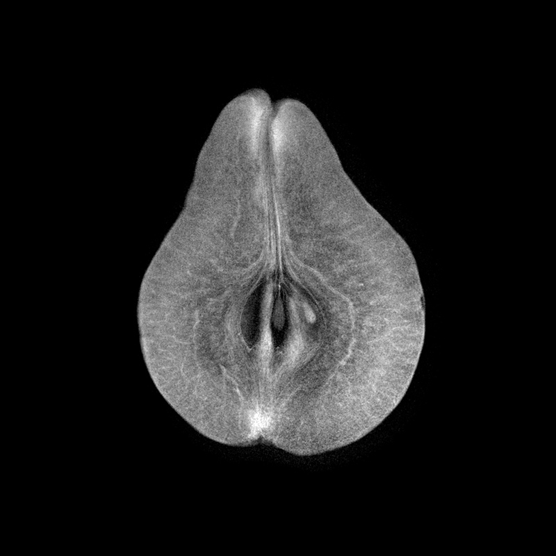
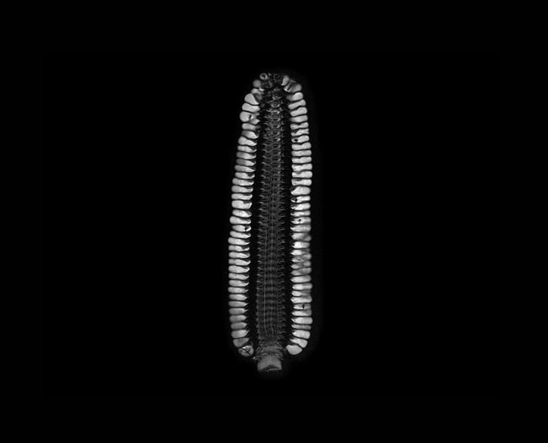
Images created with MRI
|
|

|
|
|
|
|
|
|


Back